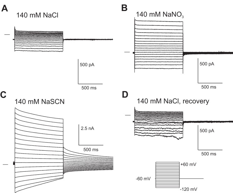
Fig. 1.
SCN− currents in isolated mouse retinal pigment epithelial (RPE) cells. Representative whole cell currents were recorded from the same RPE cell. The cell was dialyzed with a N-methyl-d-glucamine (NMDG)-Cl based K+-free internal solution and superfused with a solution containing 140 mM NaCl (A), 140mM NaNO3 (B), or 140 mM NaSCN (C). The effect of SCN− was completely reversible (D). The family of currents in D was obtained after 10 min of perfusion with 140 mM NaCl solution to ensure the complete removal of SCN−. Currents were elicited using a voltage step protocol consisting of 1-s steps, from −120 to +60 mV in intervals of 10 mV, from a holding potential of −60 mV. Horizontal lines to the left of the current traces indicate the zero-current level.

