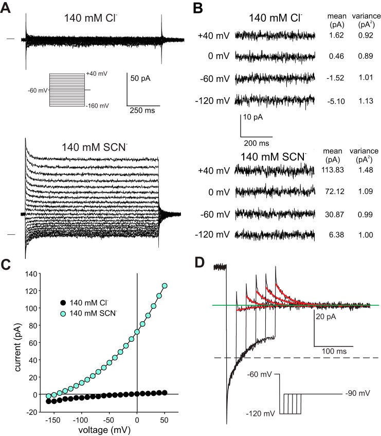
Fig. 9.
Macroscopic anion currents recorded from excised outside-out patches of basolateral membrane. A: representative current recordings from an outside-out basolateral membrane patch evoked by voltage steps in the range −160 to +40 mV from a holding potential of −60 mV in the presence of 140 mM external NaCl (top) or 140 mM external NaSCN (bottom). The horizontal line to the left of the traces indicates the zero-current level. The pipette solution contained 140 mM NMDG-Cl. Aside from brief transients that likely reflect the accumulation or depletion of SCN− on the cytoplasmic side of the patch, currents were time independent. B: subset of traces shown in A presented at higher gain. Shown to the right of each trace are the current mean and variance. Current noise in SCN− (as measured by variance) was similar to that in Cl− except at +40 mV, where it doubled. C: current-voltage (I-V) plots of steady-state currents recorded in the presence of 140 mM NaCl or NaSCN in the bath. The data are from the same patch as that depicted in A. D: tail currents obtained in a different basolateral membrane patch exposed to 140 mM external SCN−. Red traces are single exponential fits to the current records. Currents were elicited by stepping the voltage to −90 mV after hyperpolarizing the membrane to −120 mV for different durations from a holding potential of −60 mV. The tail current was inward for the initial 25 ms hyperpolarizing voltage step but was outward for voltage steps of longer duration, indicating that a time-dependent change in reversal potential (Erev) took place. This suggests that transient currents observed in outside-out basolateral membrane patches reflect local changes in SCN− concentration on the cytoplasmic side.

