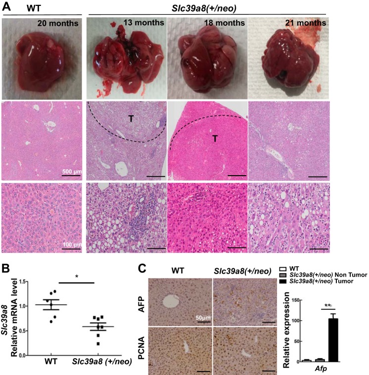
Fig. 1.
Liver pathology showing significant nodular tumors in Slc39a8(+/neo) mice. A: wild-type (WT) mouse (far left column) was 20 mo of age; Slc39a8(+/neo) mice (three right columns) were 13, 18 and 21 mo of age, respectively. Macroscopic livers (top row) and microscopic hematoxylin-eosin (H&E) staining (bottom two rows) is shown. Nodular tumors are visualized in parts or in the entire liver of Slc39a8(+/neo) mice. Out of 22 hypomorph mice, 10 were identified with similar nodular tumors. Scale bars: 500 μm (middle row); 100 μm (bottom row). B: hepatic Slc39a8 mRNA expression in WT (n = 6) and Slc39a8(+/neo) mice ages 13–21 mo (n = 7). C: typical immunohistochemistry (IHC) staining for α-fetoprotein (AFP) and proliferating cell nuclear antigen (PCNA) in WT and Slc39a8(+/neo) mice (20 mo of age) (top). Bottom: quantitative PCR analysis of Afp in WT and in nontumor portion and tumor of Slc39a8(+/neo) (n = 3 for each of the three histograms). All data represent means ± SE, with 1.0 designated for WT mean expression. *P ≤ 0.05 by Student’s t-test. T, tumor.

