Fig. 3.
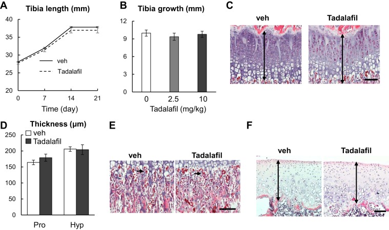
Three weeks of tadalafil treatment does not affect longitudinal skeletal growth. A: tibia length of vehicle-treated and 10 mg·kg−1·day−1 tadalafil-treated rats was measured weekly by in vivo microcomputed tomography. n = 10 rats/group. B: length of tibial in rats after 3 wk of vehicle, 2 mg·kg−1·day−1, and 10 mg·kg−1·day−1 tadalafil treatments. n = 10 rats/group. C: representative H&E staining of growth plate from vehicle group and 10 mg·kg−1·day−1 tadalafil group. Double-arrowed lines point to the growth plate. D: thickness of the proliferative zone (Pro) and hypertrophic zone (Hyp) of the growth plate was quantified. n = 5 rats/group. E: H&E staining of chondral-ossification junction of vehicle-treated and 10 mg·kg−1·day−1 tadalafil-treated rats. Arrows point to erythrocytes within blood vessels that invade growth plate cartilage. F: H&E staining of tibial articular cartilage of vehicle-treated and 10 mg·kg−1·day−1 tadalafil-treated rats. Double arrowed lines point to the articular cartilage. Scale bar: 100 µm. Results were analyzed by two-way (A, D) or one-way (B) ANOVA with Bonferroni’s post hoc test for multiple comparisons. veh, vehicle.

