Fig. 1.
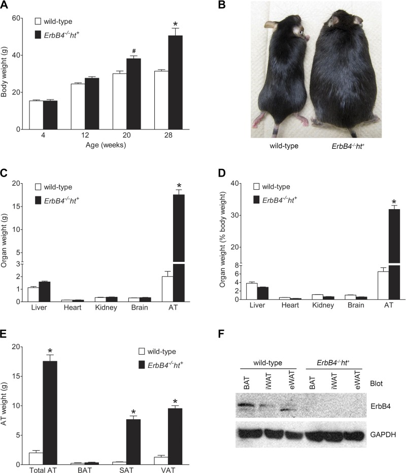
ErbB4 deletion induced obesity in mice fed the medium-fat diet (MFD). A: body weight during the MFD feeding period. Values are means ± SE (n = 9–10 in each group). #P < 0.05, *P < 0.001 vs. wild-type. B: representative images of 28-wk-old mice fed the MFD for 24 wk. C: weight of liver, heart, kidney, brain, and adipose tissue (AT) at 28 wk of age. D: organ weights in C expressed as percentage of body weight. E: weight of total AT, brown AT (BAT), subcutaneous AT (SAT), and visceral AT (VAT). F: representative image of ErbB4 expression levels in BAT, inguinal white AT (iWAT), and epididymal white AT (eWAT) in wild-type and ErbB4 deletion mice. GAPDH was used as loading control. Values are means ± SE (n = 9–10 in each group). *P < 0.001 vs. corresponding wild-type.

