Fig. 4.
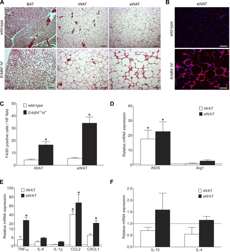
ErbB4 deletion resulted in adipocyte hypertrophy with inflammation. A: hematoxylin-eosin staining of brown adipose tissue (BAT), inguinal white adipose tissue (iWAT), and epididymal white adipose tissue (eWAT) from wild-type and ErbB4 deletion mice indicating hypertrophy with lipid accumulation in all fat depots with ErbB4 deletion compared with wild-type. Scale bars = 100 µm. B: representative image of F4/80 immunofluorescence staining. Red, F4/80; blue, DAPI. Scale bars = 100 µm. C: F4/80-positive cell quantification using ImageJ. HP, high-power. D–F: relative mRNA levels of inducible nitric oxide synthase (iNOS) and arginase 1 (Arg1), preinflammatory cytokines [TNF-α, IL-6, IL-1β, C-C motif chemokine ligand 2 (CCL2), and chemokine (C-X-C motif) ligand 1 (CXCL1)], and anti-inflammatory cytokines (IL-10 and IL-4) in iWAT and eWAT from ErbB4 deletion and wild-type (dashed line) mice. Values are means ± SE. n = 9–10. *P < 0.01 vs. wild-type.

