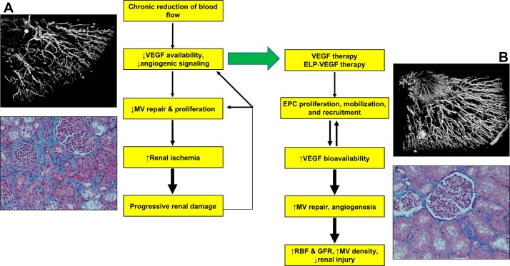
Fig. 1.
Schematic flow chart describing mechanisms of renal injury in renovascular disease and proposed mechanism of renoprotection by vascular endothelial growth factor (VEGF) therapy. Accompanied by a three-dimensional micro-CT reconstruction of the renal microcirculation (A and B, top) and a representative renal cross section showing renal fibrosis in the stenotic kidney (trichrome, ×20, A and B, bottom). MV, microvascular; ELP, elastin-like polypeptides; EPC, endothelial progenitor cells; RBF, renal blood flow; GRF, glomerular filtration rate.

