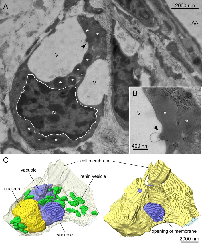
Fig. 4.
Transmission electron microscopical image (original magnification × 3,800) of a renin-producing cell of a mouse kidney perfused at 90 mmHg after 20 min of stimulation with 10 nM isoproterenol and 3 mM EGTA (A). The observed cell lies in close proximity but not adjacent to an afferent arteriole (AA). In addition, the nucleus (N), renin-containing vesicles (*), and large vacuole-like structures (V) are shown. Several renin-containing vesicles release their vesicular content into the vacuolar space (arrow). A detailed image (original magnification ×13,000) of a release of vesicular content is shown in B. 3-Dimensional reconstructions of stimulated renin-producing cells confirmed the above findings. The cell membrane is represented as a transparent layer, containing the nucleus (yellow), large vacuoles (blue), and renin-containing vesicles (green), some of them in very close proximity to the vacuoles (C, left). Additionally, the cell membrane is presented nontransparent yellow to show that the vacuoles are connected to the extracellular space (C, right).

