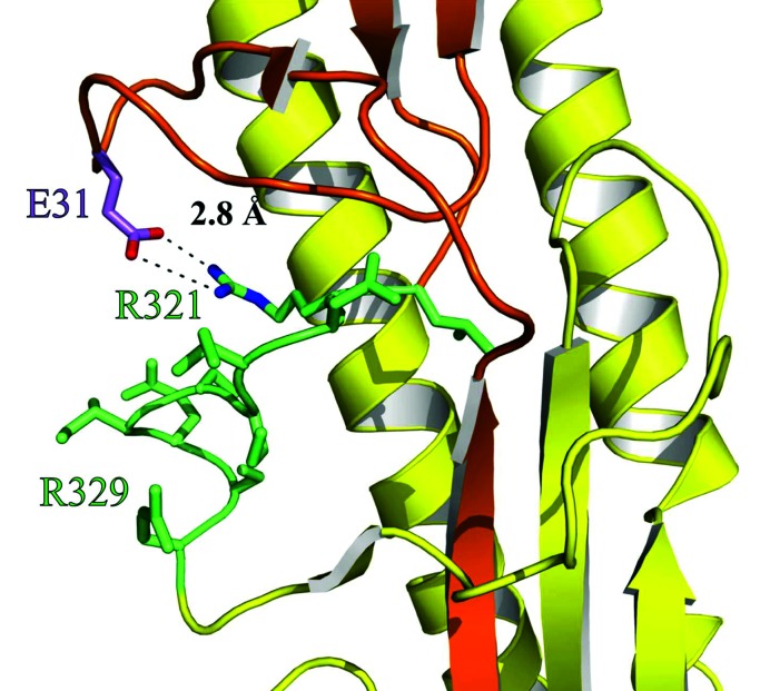
Fig. 4.
Three-dimensional structure of the cleavage site environment of haemagglutinin H1 suggests interactions with distant amino acids. Details of the haemagglutinin crystal structure from the H1N1 1918 ‘Spanish flu’ virus (PDB ID: 1RD8; [16]) showing HA1 in orange, HA2 in yellow and the HA-loop (amino acids L320-R329) in green. Residues of the HA-loop including R321 (green) and the adjacent amino acid E31 (purple) are shown as sticks. Additionally, for E31 and R321, nitrogen and oxygen atoms are coloured in blue and red, respectively, and the salt bridge is depicted as the dashed line. The figure was generated with PyMOL [37].

