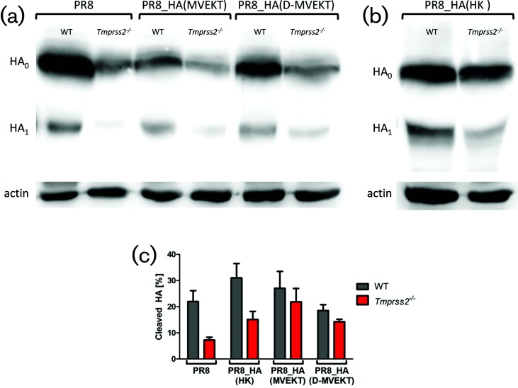
Fig. 8.
The HA of PR8_HA(MVEKT) and PR8_HA(D-MVEKT) viruses was cleaved in lungs of infected Tmprss2−/− mice. Female 8–12-week-old WT and Tmprss2−/− mice were infected intranasally with 2×10⁵ f.f.u. PR8, PR8_HA(MVEKT), and PR8_HA(D-MVEKT), and PR8_HA(HK). On day 2 p.i., lungs were homogenized and total protein was quantified. For each sample, 50 µg total protein was run on a SDS-PAGE, blotted to a PVDF membrane and stained by (a) an anti-H1N1 antibody (PR8) or (b) an anti-H3N2 antibody (A/Brisbane/10/2007) and both with a HRP-conjugated goat anti-rabbit antibody. HA0: uncleaved HA, HA1: N-terminal part of cleaved HA. Detection of signals was performed using the FujiFilm LAS-3000 imaging system. Subsequently, the same membrane was incubated with mouse anti-actin, followed by incubation with HRP-conjugated horse anti-mouse IgG and visualized again. Corresponding actin bands are shown below each sample. (c) Cleaved HA is shown as the ratio of HA1*100/(HA1+HA0). Three blots derived from the same lung homogenate samples were analysed using ImageJ software and the mean was depicted ±1 SEM.

