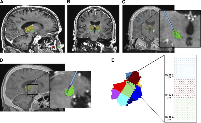
Fig. 1.
Anatomical model. A and B: preoperative MRI and 3-dimensional brain atlas fit to the patient [green volume, subthalamic nucleus (STN); yellow volume, thalamus; other nuclei not shown for clarity]. C and D: postoperative CT coregistered to the MRI and used to define the deep brain stimulation (DBS) lead location (blue shaft, pink contacts). E: STN volume divided into 9 segments that represented different regions of neuron cell density. Each voxel in the STN volume was populated with grid of points for the location of the STN neuron cell bodies.

