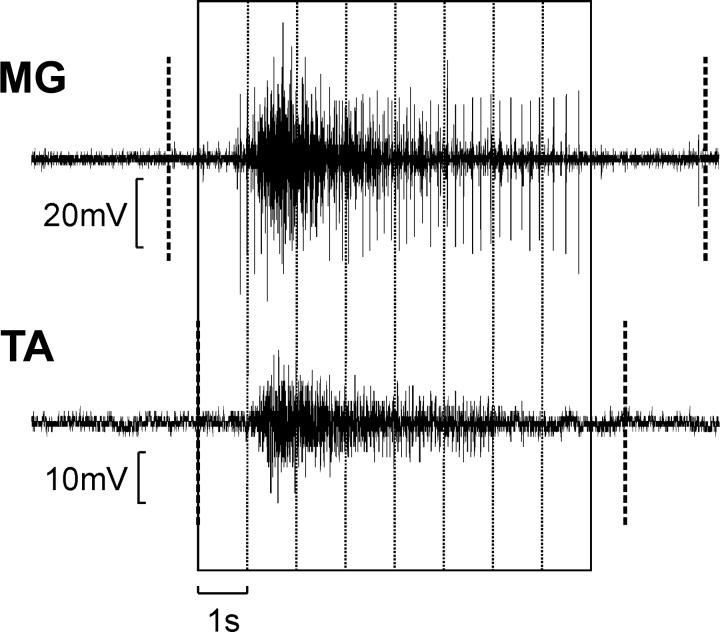
Fig. 1.
Method to calculate intermuscular coherence: example of electromyography (EMG) signals from one muscle pair (right medial gastrocnemius and tibialis anterior, MG-TA). First, the beginning and end of tonic spasms in both muscles were detected (thick dashed vertical lines). Second, data were selected only from periods where both muscles showed spasm activity simultaneously. Third, recordings were separated into 1.024-s-windows, ignoring any remaining spasm activity that could not fill a whole window. These windows were then used for the intermuscular coherence calculation.

