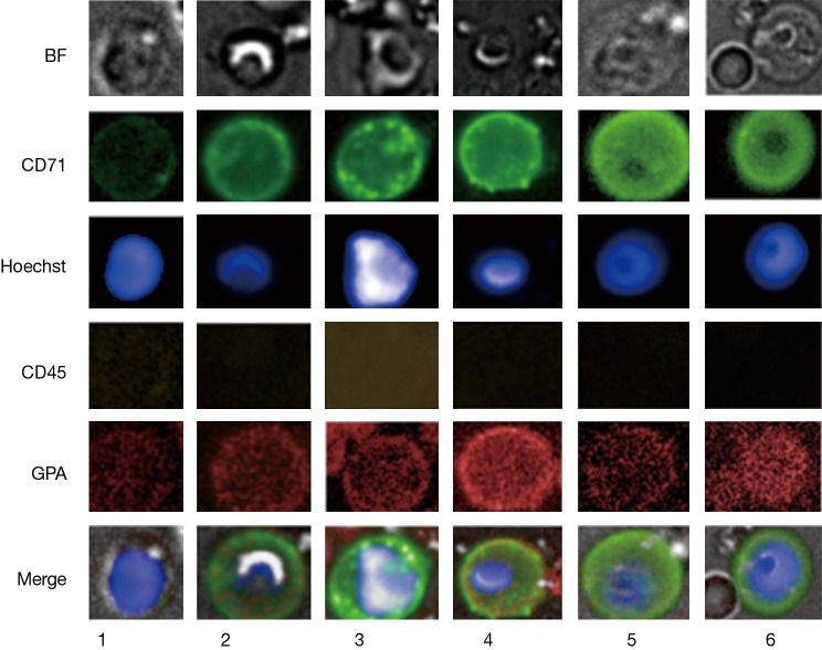
Figure 1.
Images of positive events. CD45 negative nucleated events detected by fluorescence and brightfield (BF) microscopy. A-CD71 (green), Hoechst nucleus staining (blue), a-CD45 (yellow) and a-GPA (red) immunofluorescence was used to characterize the cells. Row 1 represents small positive events measuring 8 and 5.2 µm in cell and nucleus diameter, respectively. The cell in row 2 represents a larger positive event with commonly found morphology under bright field measuring 10.7 and 6.5 µm in cell and nucleus diameter, respectively. Row 3 shows a larger cell with large atypical nucleus morphology measuring 11.5 µm in cell diameter. Row 4 represents a positive event with common bright field morphology yet larger in dimensions, measuring 13.5 and 6.5 µm in cell and nucleus diameter, respectively. Row 5 and 6 illustrate positive events with distinct bright field morphology, measuring 12.2 and 15 µm in cell diameter with nucleus diameters exceeding 7.5 µm.

