Fig. 5.
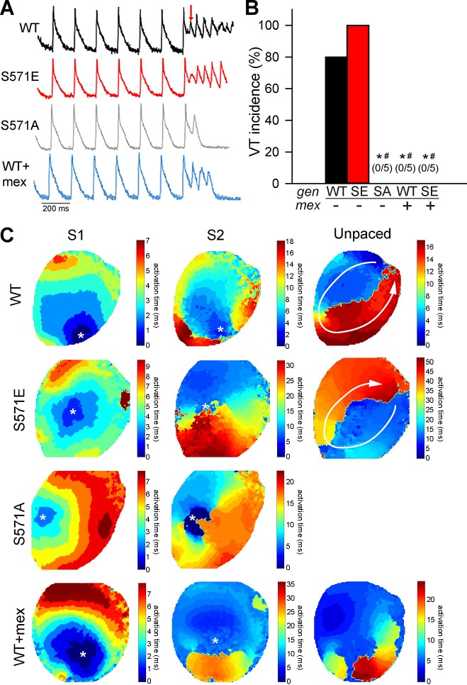
A and B: summary data and representative action potentials during the S1S2 protocol to induce arrhythmia following 5 min of reperfusion for wild-type (WT), S571 mutant (SE), and S571 mutant (SA) hearts. Data are also shown for a subset of WT and S571E hearts pretreated with mexiletine (WT + mex and S571E + mex). Cycle length of 150 ms was used for S1 pacing. The red arrow denotes the initiation of S2 protocol. VT, ventricular tachycardia. Values are means ± SE; n = 5 independent preparations for all groups. *P < 0.05 vs. WT; #P < 0.05 vs. SA. C: representative activation maps showing the spatial spread of activation after the last S1 beat, subsequent S2 beat, and first unpaced beat. WT and S571E maps demonstrate reentrant activation driving ventricular tachycardia after S2 (reentry path indicated by white arrows). Map for S571A shows conduction block near site of S2 (no unpaced activity after S2). The WT + mex map displays a single unpaced beat (no unpaced activity after the first unpaced beat). The location of pacing for S1 and S2 is indicated by a white asterisk.

