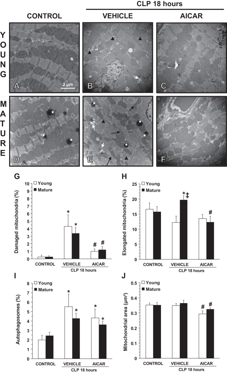
Fig. 2.
A–F: transmission electronic microscopy of cardiac cells. CLP, cecal ligation and puncture. A and D: normal cellular structure in control young (A) and mature (D) mice. B and E: structural changes in vehicle-treated young (B) and mature (E) mice at 18 h after sepsis. C and F: amelioration of mitochondrial damage in 5-amino-4-imidazole carboxamide riboside (AICAR)-treated young (C) and mature (F) mice with normal electron dense mitochondria. *Lysosomes. Black arrows show thin and elongated mitochondria. Black arrowheads show damaged mitochondria presenting translucent matrix, disrupted membrane, and cristae. White arrows show autophagic vesicles. G–J: quantification of damaged mitochondria (G), elongated mitochondria (H), autophagosomes (I), and average mitochondrial area (J) in cardiac cells as determined using NIH ImageJ software. Damaged and elongated mitochondria and autophagosomes were determined as percent total numbers of mitochondria. Data are expressed as means ± SE; n = 4 mice/group. *P < 0.05 vs. age-matched control mice; #P < 0.05 vs. age-matched mice treated with vehicle; ‡P < 0.05 vs. young mice as determined by ANOVA with Student-Newman-Keuls correction.

