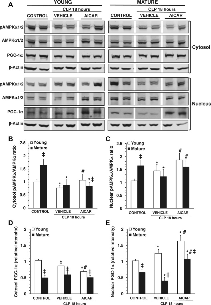
Fig. 4.
A: representative Western blot analysis of phosphorylated (p)AMP-activated protein kinase-α (AMPK-α), AMPK-α, peroxisome proliferator-activated receptor-γ coactivator (PGC)-1α, and β-actin (used as loading control protein) in heart cytosol and nuclear extracts. CLP, cecal ligation and puncture. Images were converted and adjusted in grayscale after detection by near-infrared fluorescence or chemiluminescence. B and C: image analyses of the pAMPK-α-to-AMPK-α ratio as determined by densitometry in the cytosol (B) and in the nucleus (C). D and E: image analyses of PGC-1α expression as determined by densitometry in the cytosol (D) and in the nucleus (E). Data are means ± SE of 6 control mice and 8 vehicle- or 5-amino-4-imidazole carboxamide riboside (AICAR)-treated mice for each age group and are expressed as relative intensity units. *P < 0.05 vs. baseline values of age-matched control mice; #P < 0.05 vs. age-matched mice treated with vehicle; ‡P < 0.05 vs. young mice as determined by ANOVA on ranks test.

