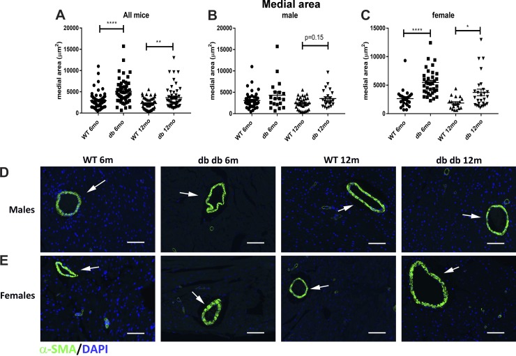
Fig. 9.
Female db/db mice show significant hypertrophy of the coronary arteriolar media. A: quantitative analysis of the picrosirius red-stained sections (shown in Fig. 8) suggested that db/db mice had a significantly higher mean arteriolar area compared with wild-type (WT) mice at 6 and 12 mo of age. B: male mice had a trend toward increased arteriolar area at 12 mo of age. C: female mice had significantly higher arteriolar area at both 6- and 12-mo time points. D and E: α-smooth muscle actin (α-SMA) immunofluorescence showed hypertrophy of the arteriolar media (arrows) in female db/db mice (n = 19–45 vessels/group for male mice, n = 19–35 vessels/group for female mice, n = 50–70 vessels/group for male + female mice). Scale bar = 50 μm. *P < 0.05, **P < 0.01, and ****P < 0.0001.

