Fig. 4.
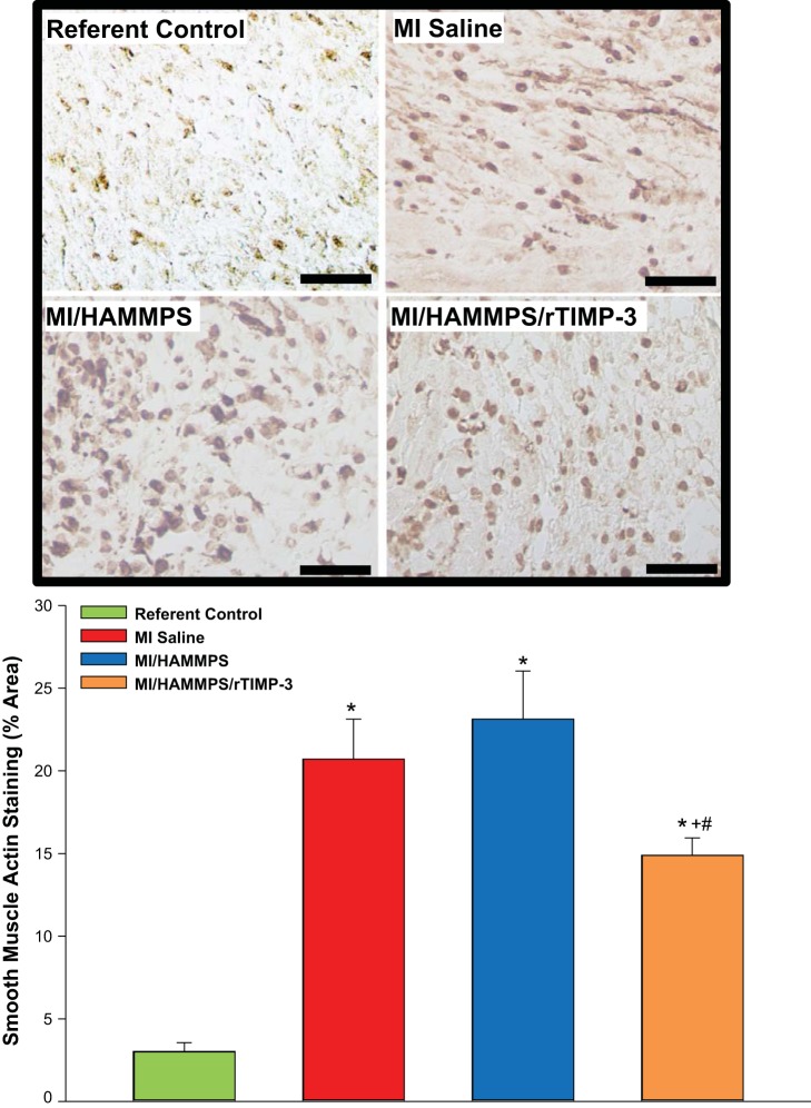
Top: representative α-smooth muscle actin (α-SMA) histochemical staining of left ventricular (LV) sections from the myocardial infarction (MI) region for all treatment groups and the corresponding LV region for referent controls. Within referent control LV sections, positive α-SMA staining was primarily localized to the vasculature, but within the MI region, it was most strongly associated with αSMA-positive cells, putatively myofibroblasts. Robust α-SMA staining was observed within the MI region for the MI/saline and MI/matrix metalloproteinase (MMP)-sensitive hyaluronic acid (HA) gel (HAMMPS) groups but appeared reduced within the MI/HAMMPS/recombinant tissue inhibitor of metalloproteinase (rTIMP)-3 group. Bottom: morphometric analysis for α-SMA staining density was performed, and a significant increase occurred in all MI groups compared with referent control values but was reduced in the MI/HAMMPS/rTIMP-3 group. Bar = 40 μm. *P < 0.05 vs. referent control; +P < 0.05 vs. MI/saline; #P < 0.05 vs. HAMMPS.

