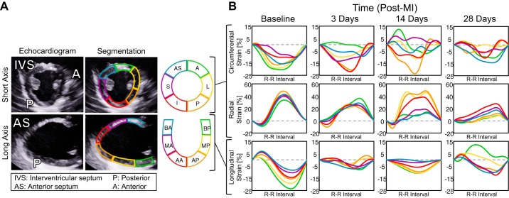
Fig. 1.
Speckle-tracking echocardiography enables quantification of myocardial segmental strain. A: representative echocardiographic images obtained 28 days post-myocardial infarction (post-MI) (at end diastole) divided into six anatomic zones for speckle tracking. B: representative segmental strain curves of the left ventricular (LV) midwall over one cardiac cycle at 28 days post-MI in the circumferential, radial, and longitudinal directions. AS, anterior septum; S, septal; I, inferior; P, posterior; L, lateral; A, anterior; BA, basal anterior septum; MA, mid anterior septum; AA, apical anterior septum; BP, basal posterior; MP, mid posterior; AP, apical posterior.

