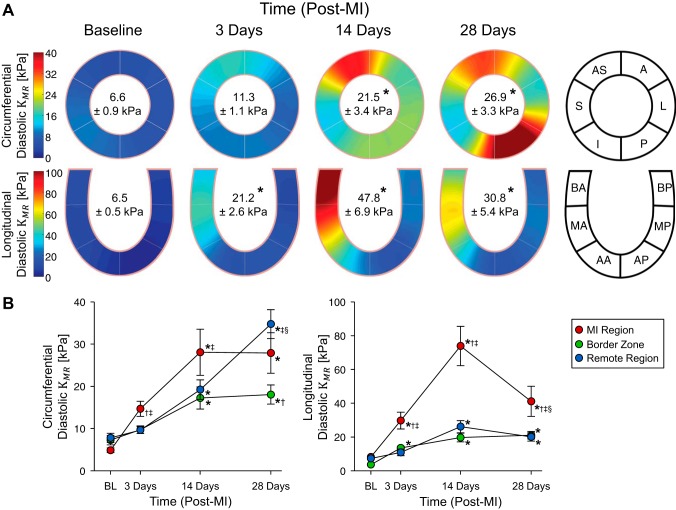
Fig. 7.
Diastolic myocardial stiffness. A: spatial maps of diastolic myocardial stiffness in the circumferential and longitudinal directions at baseline and at 3, 14, and 28 days post-myocardial infarction (post-MI). Inscribed values indicate the myocardial stiffness reported as means ± SE. B: region-specific change in myocardial stiffness post-MI. *P < 0.05 vs. the respective baseline value; †P < 0.05 vs. the respective remote region value; ‡P < 0.05 vs. the respective border zone value; §P < 0.05 vs. the respective 14-day post-MI value (n = 8, two-way ANOVA with post hoc least-significant-difference comparisons). AS, anterior septum; S, septal; I, inferior; P, posterior; L, lateral; A, anterior; BA, basal anterior septum; MA, mid anterior septum; AA, apical anterior septum; BP, basal posterior; MP, mid posterior; AP, apical posterior.

