
Figure 4.
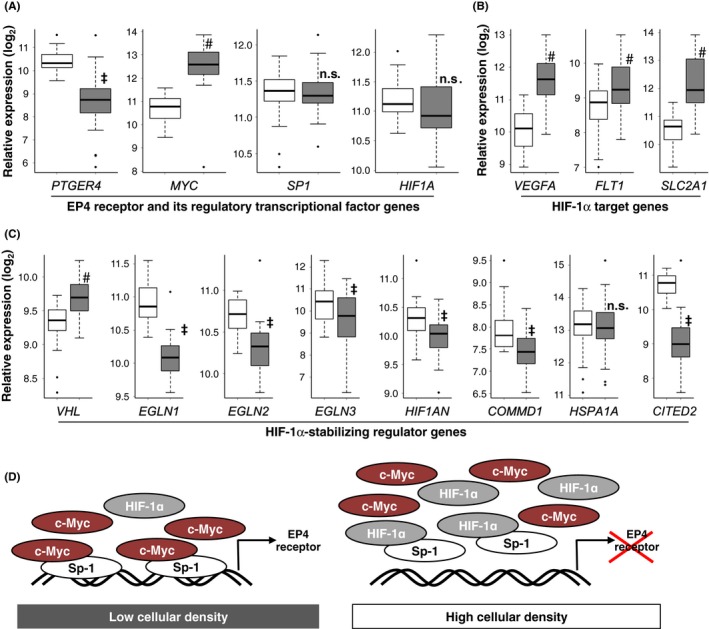
EP4 receptor and HIF1α‐related gene expression in human colon and rectal cancer tissues analyzed using the TCGA database. (A, B, C) Comparison of the mRNA expression of EP4 receptors (PTGER4), c‐Myc (MYC), Sp‐1 (SP1), and HIF‐1α (HIF1A) (A), HIF‐1α target mRNAs such as VEGF‐A165 (VEGFA), VEGFR‐1 (FLT1), and GLUT‐1 (SLC2A1) (B), and the factors involved in the stabilization of HIF‐1α such as von Hippel‐Lindau (VHL), prolyl hydroxylase domain‐containing enzymes (PHDs; also known as Egl‐9 family hypoxia‐inducible factors [EGLNs]), factor‐inhibiting HIF‐1α (FIH1; also known as HIF‐1α inhibitor [HIF1AN]), copper metabolism domain‐containing 1 (COMMD1), heat shock protein (HSP) 70 proteins, such as HSPA1A, and cAMP response element‐binding protein‐binding protein (CBP)/p300‐interacting transactivator 2 (CITED2) (C), between cancer tissues (gray boxes) and paired noncancer tissues (white boxes). & P < 0.05, the Mann‐Whitney U‐test, significantly lower than noncancer tissues. # P < 0.05, the Mann‐Whitney U‐test, significantly higher than noncancer tissues. n.s.; not significant (D) Schematic models depict the c‐Myc‐Sp‐1 complex‐mediated transcriptional activation of EP4 receptors in low cellular density‐cultured HCA‐7 cells. In high cellular density‐cultured HCA‐7 cells, increased HIF‐1α competes with and displaces c‐Myc for Sp‐1 binding and followed by pulling Sp‐1 out from its binding site, resulting in the down‐regulation of EP4 receptor transcriptional activation