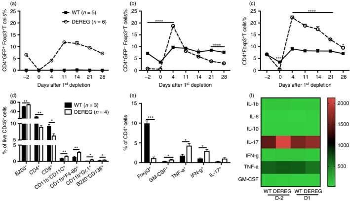
Figure 1.

Transient ablation of regulatory T (Treg) cells leads to a pro‐inflammatory milieu. Diphtheria toxin (DT) was administered in DEREG and wild‐type (WT) littermate control mice for two consecutive days, that is day 2 and 1. The frequencies of CD4+ GFP+ Foxp3+ T cells (a), CD4+ GFP− Foxp3+ T cells (b), and CD4+ Foxp3+ T cells (c) in blood were followed for 28 days. The frequencies of different immune cell populations (d) and different cytokine‐producing CD4+ T cells (e) in the spleen 1 day after the first administration of DT were determined by flow cytometry. Sera samples on day 2 and day 1 were subjected to cytokine‐plex analysis by luminex immunoassay (f). The scale indicates the concentration of cytokines (pg/ml). Error bars represent mean ± SEM and significant differences between DEREG and WT mice were determined by unpaired t‐test.
