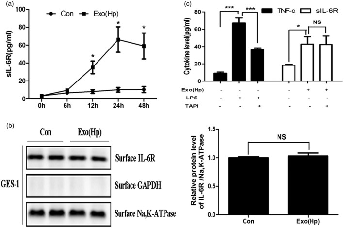
Figure 3.

Helicobacter pylori (Hp) exosomes activate sIL‐6R expression via differential splicing in gastric epithelial cells (GES)‐1 cells. (A) The levels of secreted soluble interleukin‐6 receptor (sIL‐6R) were determined using enzyme‐linked immunosorbent assay (ELISA) in GES‐1 cells at the indicated times. Con = group treated with serum exosomes from healthy volunteers. Exo(Hp) = group treated with serum exosomes from chronic gastritis patients infected with H. pylori. *P < 0·05. (b) Detection of membrane‐bound IL‐6R in GES‐1 cells following exosome treatment by Western blotting; n.s. = not significant. (c) Left: GES‐1 cells were incubated with or without TAPI (20 nM) for 8 h and treated with lipopolysaccharide (LPS) for 12 h. The protein levels of tumour necrosis factor (TNF)‐α in the culture supernatants were measured using ELISA. Right: GES‐1 cells were incubated with or without TAPI (20 nM) for 8 h and treated with exosomes for 24 h. The levels of sIL‐6R were measured using ELISA. TAPI = TNF‐α protease inhibitor. *P < 0·05, ***P < 0·001; n.s. = not significant.
