
Figure 1.
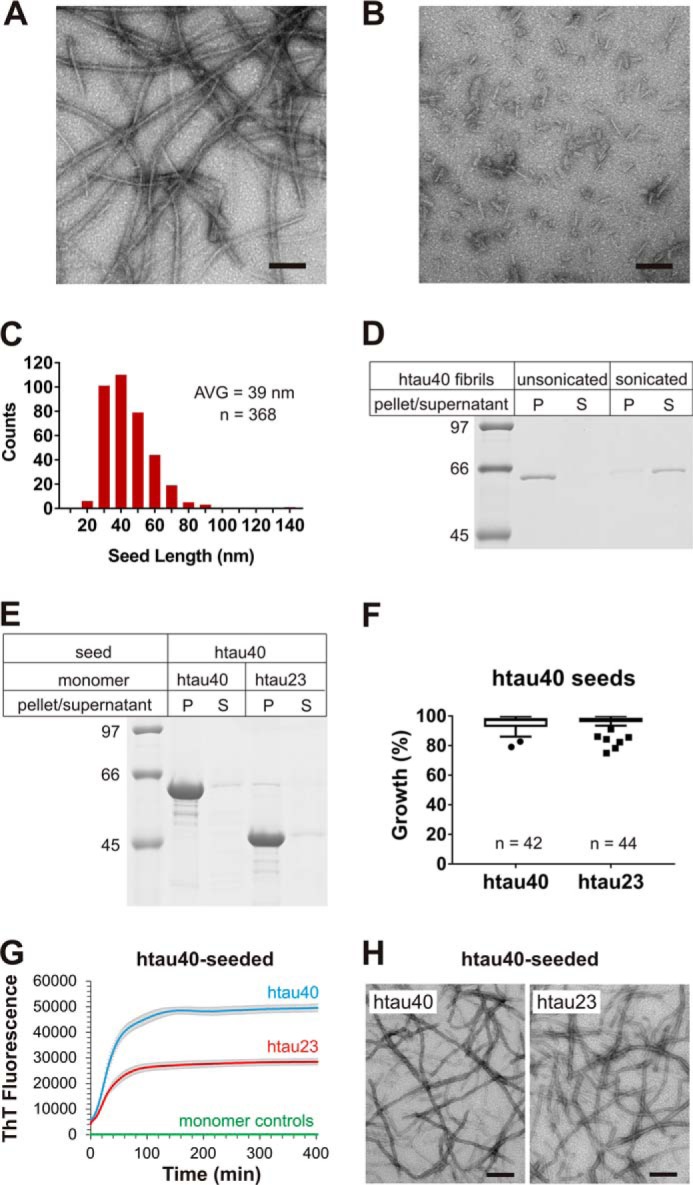
Seeding properties of htau40 fibrils. A and B, htau40 fibrils formed in the presence of heparin were analyzed by EM before (A) and after (B) sonication. Scale bars, 100 nm. C, the size distribution of sonicated fibrils reveals an average (AVG) seed length of 39 nm. D, sonicated and unsonicated fibrils were sedimented by centrifugation and analyzed by SDS–PAGE and Coomassie staining. Equivalent amounts of pellets and supernatants were loaded onto the gel. P, pellet; S, supernatant. htau40 seeds (10% monomer equivalents) were mixed with 10 μm Tau monomers and allowed to grow for 20–24 h at 37 °C. Fibrils were sedimented by ultracentrifugation. E, monomer incorporation was assessed by SDS–PAGE and Coomassie staining. F, quantitative analysis of seeding properties using gel densitometry depicted as box-and-whisker plot (Tukey method). n, number of biological replicates. G, ThT fluorescence measurements of htau40 (blue trace) and htau23 (red trace) growth onto htau40 seeds (10 μm monomers, 5% seeds) in triplicate. The t½ values for these reactions were 27 and 25 min, respectively. Triplicate ThT measurements of htau23 and htau40 monomers in the absence of seeds (overlapping green traces). Error bars represent means ± S.D. H, negative stain EM images of htau40 (left panel) and htau23 (right panel) fibrils after seeding. Scale bars, 200 nm. Collectively, the results demonstrate growth of htau23 and htau40 monomers onto htau40 seeds.