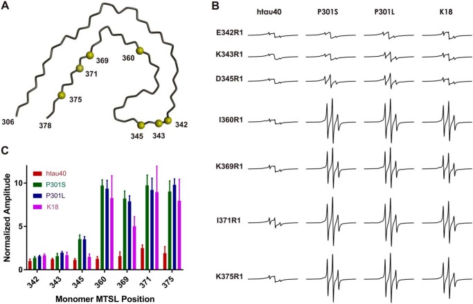
Figure 8.
EPR analysis of fibrils seeded with WT, mutant, and truncated Tau. Seven single cysteine mutants of htau40 were first labeled with the paramagnetic spin label MTSL, then combined with Tau fibril seeds (htau40, P301S, P301L, and K18), and incubated for 20–24 h at 37 °C. A, all cysteines were located at positions that in the paired helical filament point to the aqueous exterior. The depicted model represents the protein backbone of a single Tau layer. In the filament thousands of these layers are stacked on top of each other. The model utilizes PDB accession number 5O3L. Yellow spheres depict Cα carbons at the sites of cysteine substitution. B, representative CW EPR spectra of Tau fibrils collected at X-band. The horizontal row on top signifies the type of seeds that was used. The vertical column on the left identifies the spin labeled htau40 monomer that was grown onto the seed. R1, spin-labeled cysteine. All spectra are normalized to the same number of spins. Scan width, 150 G; modulation, 3 G; incident microwave power, 12 milliwatt. C, plot of the signal amplitudes. The results are represented as the means ± S.D. using three biological replicates. The data highlight differences in the core structures of the fibrils.

