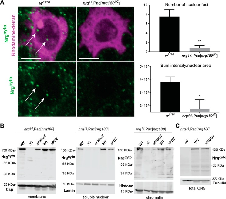
Figure 2.
Subcellular localization of Drosophila Nrg in WT and Nrg mutants. A, subcellular localization of Nrg180 in the WT (w1118) and nrg14;Pac[nrg180ΔC] negative control flies with Nrg180cyto labeling. WT and negative control animals were immunohistochemically processed in parallel and scanned with the same settings. Shown is the image of single confocal slice through the nucleus of the giant fiber soma dye filled with rhodamine-dextran (magenta), which is preferentially excluded from the nucleus. Nrg180 (green) labeled puncta were seen in the cytoplasm and the nucleus (arrows) of w1118 flies. Scale bar, 5 μm. Right, quantification of Nrg180 labeling with Nrg180cyto antibodies in the nucleus. The number of puncta (p < 0.003, two-tailed Student's t test) and the sum of fluorescent intensity levels (p < 0.026, two-tailed Student's t test) normalized to the total nuclear areas is significantly different in WT (n = 6) and negative control (n = 5) animals. B, Western blots of the membrane (10 heads), soluble nuclear (100 heads), and chromatin (100 heads) fractions (Subcellular Protein Fractionation Kit) of WT (nrg14;Pac[WT]), negative controls (nrg14;Pac[nrg180ΔC]), and Nrg pacman mutants (nrg14;Pac[nrg180ΔPDZ], nrg14;Pac[nrg180ΔFIGQY]) lacking the FIGQY or C-terminal PDZ motifs probed with Nrg180cyto antibodies. Anti-Csp, anti-lamin, and anti-histone were used as controls for the membrane, soluble nuclear, and chromatin fractions, respectively. C, Western blotting of total protein extracts of 20 heads from nrg14;Pac[nrg180ΔC], nrg14;Pac[WT], and nrg14;Pac[nrg180ΔFIGQY] animals probed with Nrg180cyto antibodies. Anti-tubulin served as a loading control. Error bars represent standard error of the mean. Two-tailed Student′s t test was used to determine statistically significant differences (*, p ≤ 0.05, **, p < 0.01).

