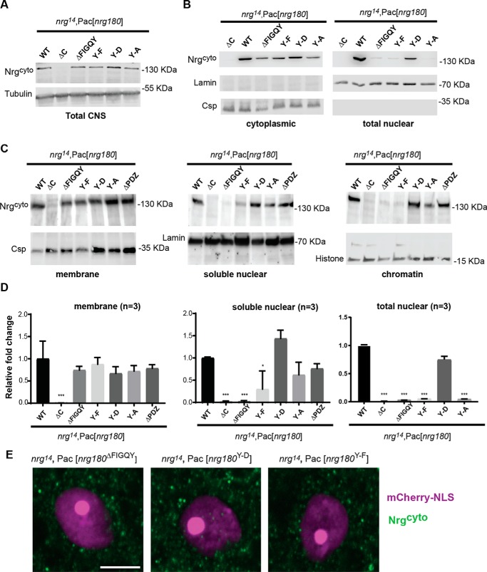
Figure 3.
Subcellular localization of Nrg FIGQY mutants. A, Western blotting of 10 heads of WT controls (nrg14;Pac[WT]) and Nrg loss-of-function mutants (nrg14;Pac[nrg180ΔC], nrg14;Pac[nrg180ΔFIGQY], nrg14;Pac[nrg180Y-A], nrg14;Pac[nrg180Y-D], and nrg14;Pac[nrg180Y-F]). Tubulin served as a loading control. B, Western blots of cytoplasmic and nuclear fractions of WT controls and Nrg180 mutants using Thermo Fisher Scientific NE-PER® kit. The cytoplasmic fraction of ∼20 brains and the nuclear fraction of ∼130 brains were loaded per lane. Anti-Csp and anti-lamin labeling was used as loading control. C, Western blotting of the membrane (10 heads), soluble nuclear (100 heads), and chromatin (100 heads) fractions from WT controls (nrg14;Pac[WT]) and Nrg loss-of-function mutants (nrg14; Pac[nrg180ΔC], nrg14;Pac[nrg180ΔFIGQY], nrg14;Pac[nrg180Y-A], nrg14;Pac[nrg180Y-D], nrg14;Pac[nrg180Y-F], and nrg14;Pac[nrg180ΔPDZ]) probed with Nrg180cyto antibodies. Anti-Csp, anti-lamin, and anti-histone served as controls for the membrane, soluble nuclear, and the chromatin fractions, respectively. D, quantitative densitometry analysis of three independent Western blots of membrane (Subcellular Protein Fractionation Kit), soluble nuclear (Subcellular Protein Fractionation Kit), and nuclear (Thermo Fisher Scientific NE-PER®) fractions from WT controls (nrg14;Pac[WT]) and Nrg180 mutants probed with Nrg180cyto antibodies. Error bars, S.E. Statistical significance was assessed using Student's t test (*, p ≤ 0.05; **, p < 0.01; ***, p < 0.001). E, GF somas of nrg14;Pac[nrg180ΔFIGQY], nrg14;Pac[nrg180Y-D], and nrg14;Pac[nrg180Y-F] animals were labeled by expression of mCherry-NLS (magenta) with the R68A06-Gal4 line. Images of single confocal slices through the nucleus of the giant fiber somas labeled with Nrg180cyto (green) are shown. Scale bar, 5 μm.

