Figure 2.
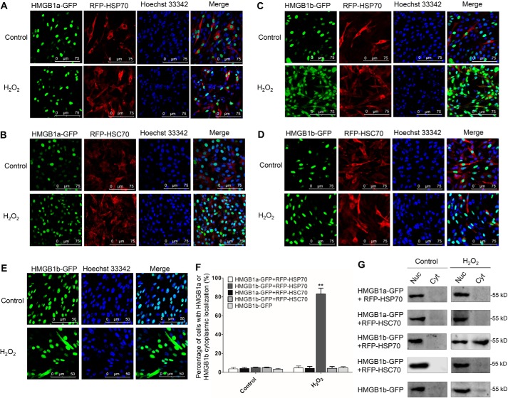
HSP70 specifically promotes H2O2-induced nucleocytoplasmic translocation of HMGB1b. A–E, CIK cells stably expressing HMGB1a-GFP and RFP-HSP70 (A), HMGB1a-GFP and RFP-HSC70 (B), HMGB1b-GFP and RFP-HSP70 (C), HMGB1b-GFP and RFP-HSC70 (D), or HMGB1b-GFP (E) were seeded on microscope coverglasses in 12-well plates for 24 h at about 90% confluence and treated with H2O2 at a nontoxic dose (0.15 mm) or PBS for 36 h. Then the cells were fixed with 4% (v/v) paraformaldehyde and stained with Hoechst 33342. Subsequently, all samples were visualized using a confocal microscope. F, counting analysis of cells with HMGB1a or HMGB1b cytoplasmic localization. Error bars indicate S.D. (n = 4). **, p < 0.01. G, nucleocytoplasmic distribution of HMGBs was examined by WB. The indicated cells were treated with H2O2 or PBS for 36 h in 10-cm dishes. Then the nuclear (Nuc) and cytoplasmic (Cyt) proteins were extracted. The protein levels of HMGB1b-GFP in different cell fractions were examined by IB analysis with GFP Ab.

