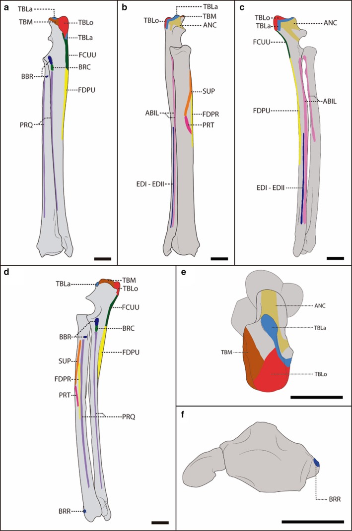
Figure 3.

Schematic representation of the radius and ulna of a female, adult specimen of Lycalopex gymnocercus (8576) illustrating muscle insertions. Caudal (a), cranial (b), lateral (c), medial (d), proximal (e) and distal (f) views, with details on the areas of muscle insertion of the intrinsic muscles: ABIL, abductor digiti I longus; ANC, anconeus; BBR, biceps brachii; BRC, brachialis; BRR, brachioradialis; TBLa, triceps brachii caput lateralis; TBLo, triceps brachii caput longum; TBM, triceps brachii caput medialis; FCUU, flexor carpi ulnaris caput ulnare; FDPR, flexor digitorum profundus caput radiale; FDPU, flexor digitorum profundus caput ulnare; EDI‐EDII, extensor digiti I and II; PRQ, pronator quadrates; PRT, pronator teres; SUP, supinator. Scale bar: 10 mm.
