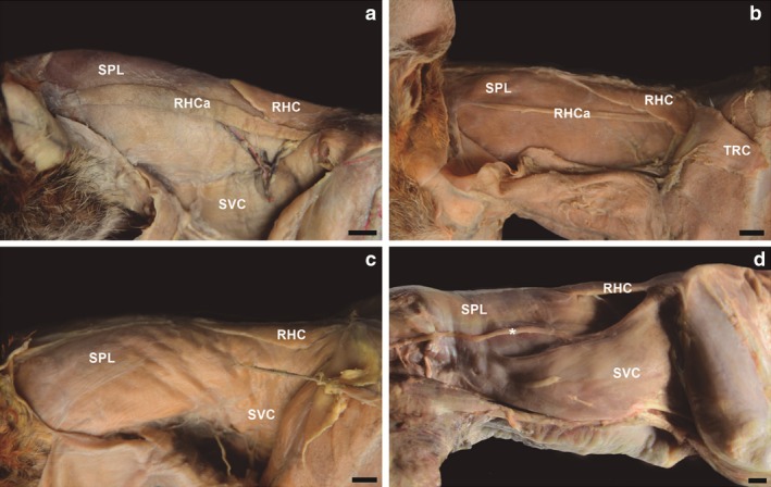
Figure 5.

Photomacrographs of the muscles in the lateral cervical region of four adult specimens of Lycalopex gymnocercus. The most common presentation was a well‐developed m. rhomboideus capitis (a). However, variations with little developed (b) or absent (c) m. rhomboideus capitis were also observed. Another variation was a thin muscle strip (*) apparent in m. serratus ventralis cervicis in specimens that did not show m. rhomboideus capitis. SVC, m. serratus ventralis cervicis; RHC, rhomboideus cervicis; RHCa, m. rhomboideus capitis; SPL, m. splenius; TRC, m. trapezius pars cervicalis. Scale bar: 10 mm.
