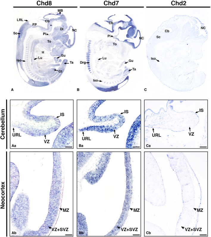
Figure 1.

Distinct Chd8, Chd7 and Chd2 expression patterns at E12.5. In situ hybridisation on sagittal sections of mouse embryos at developmental stage E12.5 using antisense riboprobes to detect Chd8, Chd7 and Chd2 mRNA, anterior to the right (A–C). Gene expression is indicated by purple/blue staining. Note the widespread Chd8 expression in most embryonic tissues (A), the high, localised expression of Chd7, specifically in the developing nervous system (B), and very low, widespread expression of Chd2 (C). High‐magnification images of the developing cerebellum (Aa–Ca) demonstrate the presence of Chd8 (Aa) and Chd7 (Ba) transcripts throughout the neuroepithelium, with little Chd2 expression evident (Ca). High‐magnification images through the neocortex show widespread Chd8 expression (Ab), note that Chd7 expression tends to be higher on the ventricular side (Bb) and that there is little discernible Chd2 expression (Cb). Other regions with relatively strong signals were the nasal epithelium, tail, genital tubercle, intersomitic mesoderm, spinal cord, mid brain, diencephalon, tongue and pituitary. Scale bars: 100 μm. Cb, cerebellum; Di, diencephalon; Drg, dorsal root ganglia; FP, floor plate; Gt, genital tubercle; Gu, gut; H, heart; Is, isthmus; Iso, intersomitic mesoderm; Lu, lungs; LRL, lower rhombic lip; MB, midbrain; MZ, molecular zone; NC, neocortex; Pi, pituitary; Sc, spinal cord; SVZ, subventricular zone; Ta, tail; To, tongue; URL, upper rhombic lip; VZ, ventricular zone.
