Figure 3.

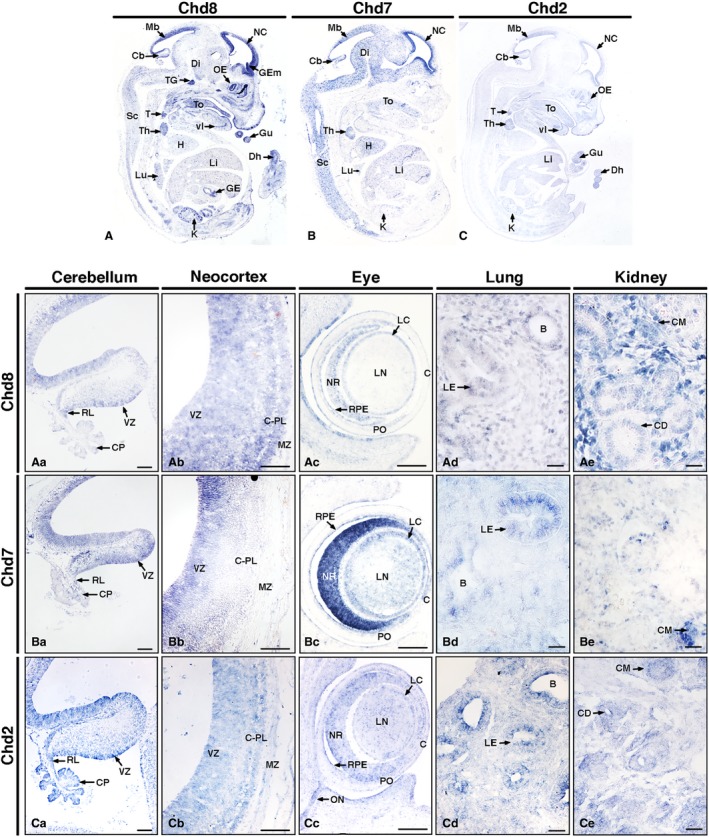
Distinct nervous system and organ‐specific expression patterns of Chd8, Chd7 and Chd2 in E14.5 mouse embryos. In situ hybridisation on sagittal sections of E14.5 mouse embryos (A–C), anterior to the right. Note distinct Chd8, Chd7 and Chd2 expression patterns throughout the embryos with notably higher levels in the developing nervous system. Beyond the nervous system, other notable regions of expression included various organs and glands, for example the thymus and thyroid, heart and kidneys. High‐magnification images (Aa–Ce) revealed specific expression patterns in the cerebellum (Aa–Ca), neocortex (Ab–Cb), eye (Ac–Cc), lung (Ad–Cd) and kidney (Ae–Ce). Scale bars: 100 μm. B, bronchus; C, cornea; Cb, cerebellum; CD, collecting duct; CM, condensing mesenchyme; CP, choroid plexus; C‐PL, cortical plate; Di, diencephalon; Dh, digit of hindlimb; GE, gastric epithelium; GEm, ganglionic eminence; Gu, gut; H, heart; K, kidney; LC, lens capsule; LE, lung epithelium; Li, liver; LN, lens; Lu, lung; Mb, mid brain roof plate; MZ, marginal zone; NC, neocortex; NR, neural retina; OE, olfactory epithelium; ON, optic nerve and surrounding structures; PO, pre‐optic cup; RL, rhombic lip; RPE, retinal pigmented epithelium; Sc, spinal cord; T, thyroid; To, tongue; Ta, tail; Th, thymus; TG, trigeminal ganglion; vI, ventral incisor; VZ, ventricular zone.
