
Figure 3.
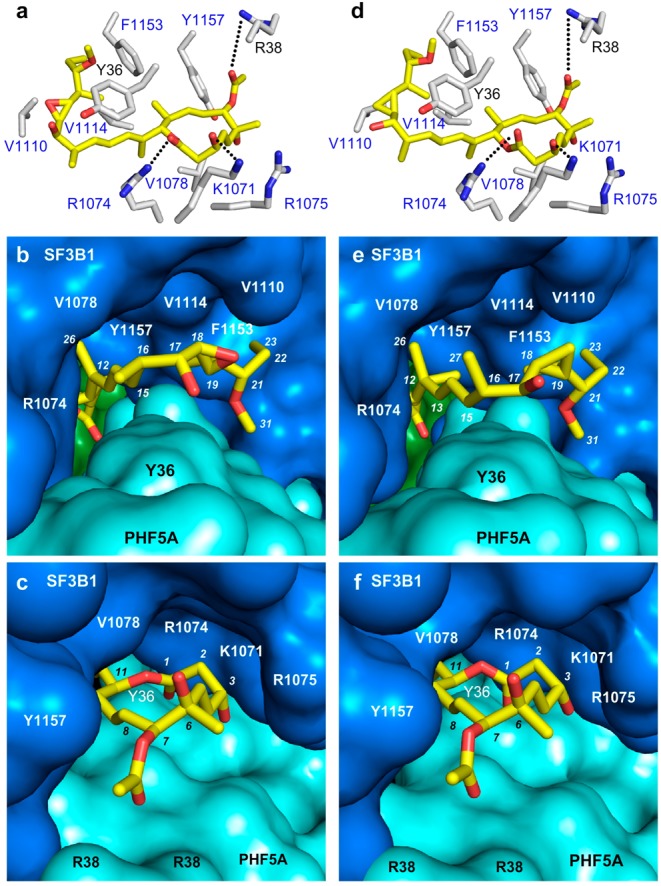
Co-crystal structures of (a–c) FD-895 (1a) and (d–f) cyclopropane 1c within the SF3B core. (a,d) Side chains of residues within 6 Å of the SPLM (yellow) are labeled in gray corresponding to SF3B1 (blue labels) and PHF5A (black labels). (b,e) “Connelly” surfaces of the SPLM binding site showing side chains of 1a and 1c occupying a pocket formed at the interface between SF3B1 (blue), PHF5A (cyan), and SF3B3 (green). (c,f) “Connelly” surfaces in the SPLM binding site depicting the macrolides of 1a and 1c positioned at the other end of this tunnel.