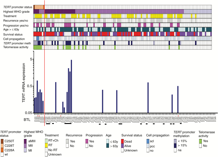
Fig. 1 .
TERT promoter status linked to clinical and cell-biological characteristics and telomere stabilization parameters in 128 meningioma samples of 110 patients. Data on mutation status, tumor grade, treatment, tumor recurrence, tumor progression, patient age, survival status, cell propagation and TERT promoter methylation as well as telomerase activity are indicated in different colors. The respective legend is outlined below the graph. aMIII, anaplastic meningioma WHO grade III; secondary anaplastic meningiomas WHO III are indicated with white asterisks; MII, atypical meningioma WHO grade II; MI, meningothelial (47%; 15/32), transitional (31%; 10/32), fibroblastic (16%; 5/32), angiomatous (6%; 2/32) meningioma WHO grade I; RT, radiotherapy; Ch, chemotherapy; scl, stable cell line; pcc, primo-cell culture; TERT promoter methylation according to the cutoff of 15%; na, not analyzed. Patients with corresponding primary and recurrent tumor samples are highlighted with horizontal black bars below the graph depicting TERT mRNA expression levels by RT-PCR. The blue asterisks in the recurrence and progression lines highlight a special case of meningioma WHO III which recurred with a gliosarcoma 14 months after the first diagnosis. The respective BTL numbers of meningioma-derived cell cultures used for the in vitro experiments are marked with red asterisks.

