Fig. 5.
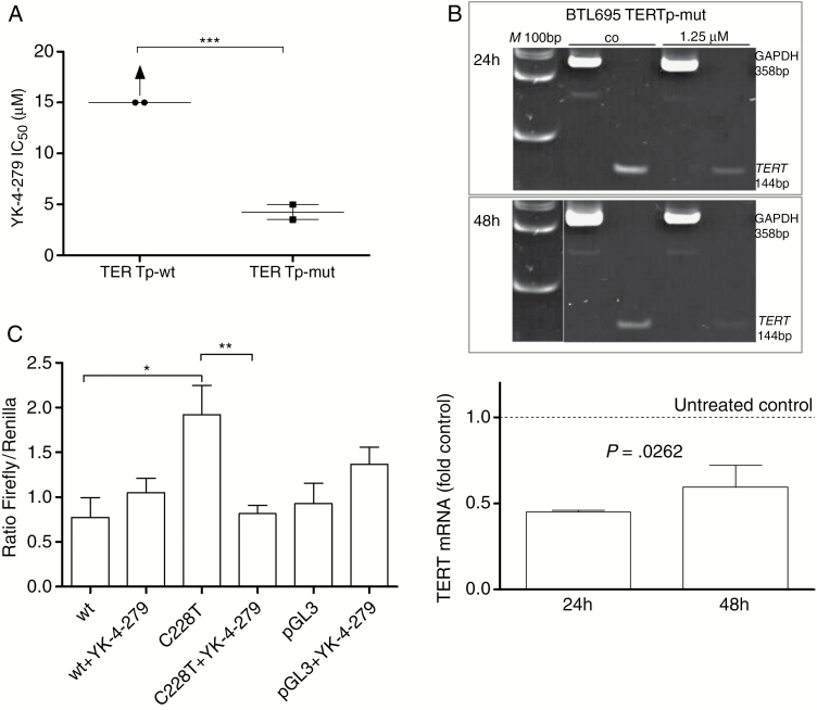
TERT promoter status and responsiveness to the ETS-factor inhibitor YK-4–279. (A) Meningioma primo-cell cultures with TERTp-wt (BTL400, BTL2282) and TERTp-mut (BTL695, BTL598) backgrounds were exposed to increasing concentrations of YK-4–279, and cell survival rates were determined by MTT assays. Mean IC50 values are given. In the case of TERTp-wt status, IC50 values were above the highest applicable YK-4–279 concentration (15 µM) but estimated at 15 µM for statistical analysis. (B) Downregulation of TERT mRNA expression analyzed by RT-PCR in the TERTp-mut BTL695 meningioma cell line after 24 and 48 h drug exposures as depicted. YK-4–279 treatment resulted in a marked decrease of TERT mRNA in the TERTp-mut BTL695 cell line (upper panel). M, 100 bp size marker. Additionally, quantitative real-time PCR was performed proving significant TERT mRNA downregulation by exposure to 1.25 µM YK-4–279. TERT mRNA expression levels are given relatively to the untreated control set as 1. Data are derived from 2 independent experiments performed in triplicates. (C) BTL695 cells were transfected with luciferase reporter constructs containing either the wild-type (wt) or the C228T TERT promoter sequence. Forty-eight hours post transfection, cells were treated with 5 µM YK-4–279 and 18 h later reporter expression was analyzed. Values are depicted as ratios firefly/Renilla. Negative control was pGL3 basic vector. Significant differences were calculated in panels A and C using Student’s t-test (***P < 0.001, **P < 0.01, *P < 0.05) and one-way ANOVA in panel B.

