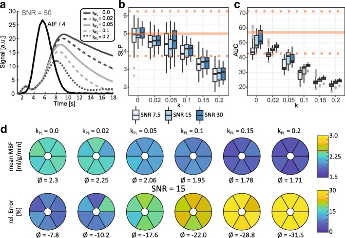
Fig. 4.
Simulation results for variations of kinetic conversion rate parameter k for SNR = 7.5, 15 and 30. Reference values from input concentration dynamics are indicated by the solid line. Dotted lines indicate the ±25% interval around the reference value. a Illustration of changes in myocardial response curve with increasing k values. Faster conversion at higher values results in a reduced pyruvate signal. Downstream lactate and Bicarbonate signal can cause signal contamination as illustrated in Fig. 2. b-c Semi-quantitative perfusion measures as function of k values analogous to Fig. 3. b Myocardial upslope (SLP). For k values ≥0.05 the SLP index is noticeably affected. c Area under the curve (AUC). d Mean absolute MBF quantification with relative error per myocardial sector for SNR = 15. Increasing conversion rates cause systematic underestimation of MBF. For values ≤0.1 the error is below 20%

