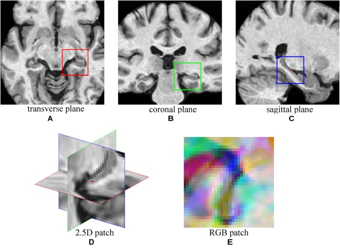
FIGURE 2.
The demonstration of 2.5D patch extraction from hippocampus region. (A–C) 2D patches extracted from transverse (red box), coronal (green box), and sagittal (blue box) plane; (D) The 2.5D patch with three patches at their spatial locations, red dot is the center of 2.5D patch; (E) Three patches are combined into RGB patch as red (red box patch), green (green box patch), and blue (blue box patch) channels.

