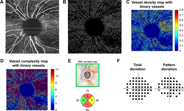
Figure 3.
Moderate-severe glaucoma case (A) 6×6 mm2 en face image. (B) Skeletonized vessel image with large vessels removed. (C) Vessel density map with binary vessels, showing areas of higher vessel density in warmer colors. (D) Vessel complexity map with binary vessels, showing areas of greater vessel branching in warmer colors. (E) Cirrus OCT RNFL deviation map (top) and RNFL thickness by quadrant (bottom). (F) Probability total (left) and pattern (right) deviation maps.
Abbreviations: I, inferior quadrant; N, nasal quadrant; OCT, optical coherence tomography; RNFL, retinal nerve fiber layer; S, superior quadrant; T, temporal quadrant.

