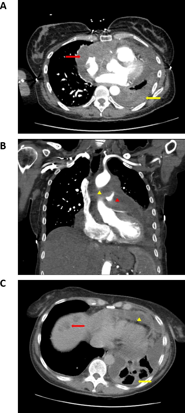
Figure 1. CT imaging of cardiac angiosarcoma prior to propranolol treatment.

(A) Baseline axial chest CT showing a large soft-tissue mass invading the right atrium and encasing the heart (red arrow). There is also nodular pleural thickening (yellow arrow) along the costal and mediastinal pleural surfaces and a small pleural effusion secondary to pleural infiltration. (B) Baseline coronal chest CT shows soft tissue mass encasing the ascending aorta (yellow arrowhead) and main pulmonary artery (yellow arrowhead). (C) Baseline axial chest CT shows circumferential pericardial thickening and effusion (yellow arrowhead) and nodular pleural thickening (yellow arrow) with loculated pleural effusion. Note small hypodense lesions in the right lobe of the liver consistent with metastases (red arrow).
