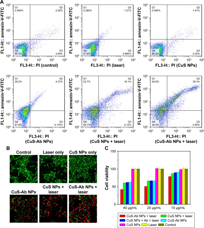
Figure 3.
Cytotoxicity and cellular uptake of CuS NPs in vitro.
Notes: (A) The cytotoxicity of NPs was evaluated by flow cytometry after co-culture with 4T1 cells. (B) Cell viability in each group was evaluated by calcein AM/PI co-staining with or without 1,064 nm laser irradiation (magnification: 40× objective; calcein AM, green indicates living cells; PI, red indicates dead cells). (C) Viability of 4T1 cells in all groups with or without CuS NPs at different concentrations in the presence or absence of irradiation. (D) Viability of HUVECs was assessed by MTT without laser irradiation. (E) Cellular uptake of CuS NPs and CuS-Ab NPs was observed by laser scanning confocal microscopy (60×, oil objective). Green indicates FITC-CuS NPs and FITC-CuS-Ab NPs; red indicates lysosomes; blue indicates cell nuclei. All data represent mean values (n=3).
Abbreviations: calcein AM, calcein-acetoxymethyl ester; CuS NP, CuS nanoparticle; CuS-Ab NP, cetuximab-modified CuS NP; FITC, fluorescein isothiocyanate; HUVEC, human umbilical vein endothelial cell; PI, propidium iodide.


