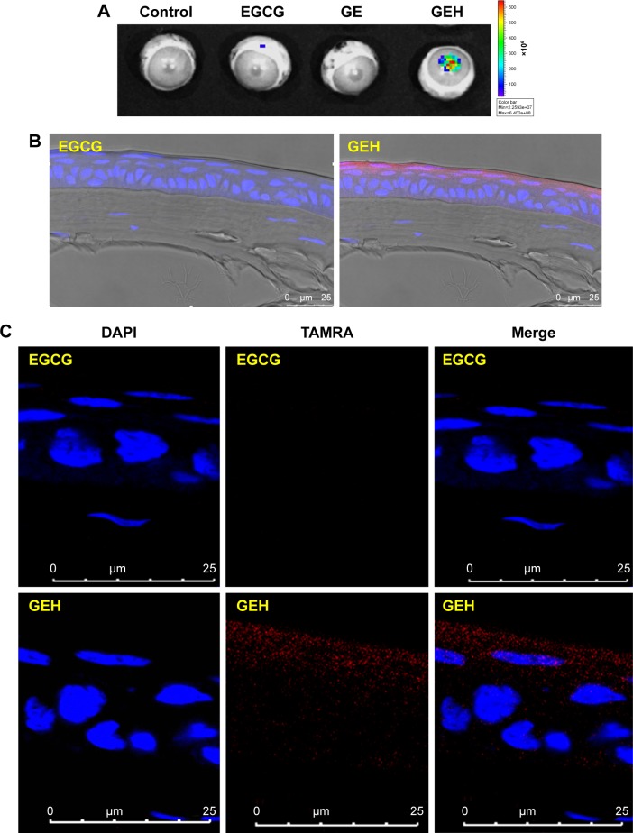
Figure 7.
NPs location and distribution in cornea.
Notes: (A) Accumulation of fluorescent particles on rabbit eye after 2 hours’ dosing with different formulations captured by in vivo imaging system. (B and C) Confocal microscopy of dye or NP distribution in corneal epithelium from a cryosection of rabbit cornea in images at (B) low and (C) high magnification (TAMRA 0.5 µg/mL).
Abbreviations: EGCG, epigallocatechin gallate; GE, gelatin–EGCG; GEH, GE with hyaluronic acid coating; NP, nanoparticle; TAMRA, tetramethylrhodamine.

