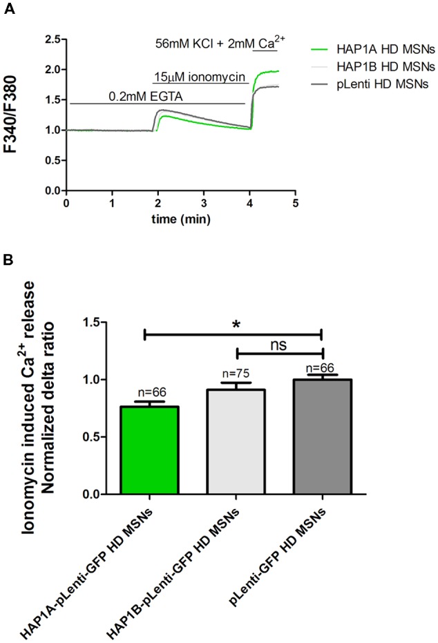
Figure 4.

Effect of HAP1A on ER Ca2+ content in YAC128 MSNs. Cultures of MSNs on DIV14 from YAC128 mice that overexpressed HAP1A-pLenti-GFP, HAP1B-pLenti-GFP, or pLenti-GFP using lentiviruses that were loaded with the Ca2+ indicator Fura-2AM. (A) The figure shows the protocol to induce ionomycin-induced Ca2+ release from the ER in YAC128 MSNs that overexpressed HAP1 isoforms using the eight-well system. YAC128 MSNs that overexpressed HAP1A isoforms were incubated in Ca2-free medium (0.1 mM EGTA), followed by 15 μM ionomycin application to induce the release of Ca2+. KCl (56 nM) in 2 mM Ca2+ was applied to distinguish neurons from glial cells. (B) Ionomycin-induced ER Ca2+ release in YAC128 MSNs that overexpressed HAP1 isoforms. The results are expressed as mean ± SEM. The number of cells is shown on the top of the bars. *p < 0.05. ns, not significant. The results were obtained from three independent MSN culture preparations.
