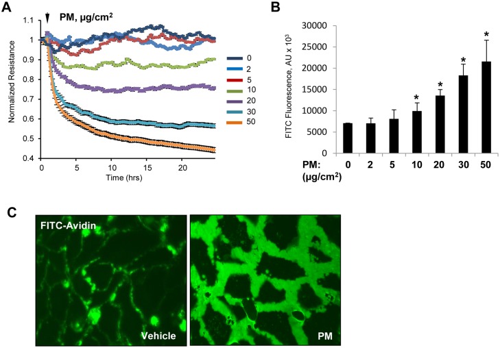
Fig 1. PM causes endothelial barrier disruption.
(A) Human pulmonary lung EC were exposed to indicated concentrations of PM and TER was measured across the cell monolayers over time. (B) Cells grown on immobilized biotinylated gelatin were exposed to PM for 4 hr and FITC-avidin (25 μg/mL) was added for 3 min. After washing unbound FITC-avidin with PBS, FITC fluorescence was determined in Victor X5 plate reader. Normalized readings are expressed as mean ± S.D.; n = 6, *p<0.05. (C) Visualization of FITC fluorescence in control or PM-treated cells (20 μg/cm2, 4 h).

