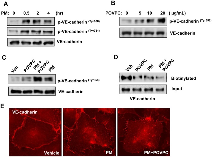
Fig 6. Tr-OxPLs augment PM-induced AJ disruption.
(A) Cells were treated with PM for indicated time periods, and phospho-VE-cadherin (Tyr 658 and Tyr 731) levels were determined by Western blotting. (B) Cells were treated with increasing concentrations of POVPC for 30 min, and cell lysates were analyzed by Western blotting to detect phospho-VE-cadherin. (C, D) Cells were challenged with low dose of POVPC (10 μg/mL) alone or in combination with PM, and phospho-VE-cadherin levels (C) or biotinylated VE-cadherin levels (D) were detected by Western blotting. The total cell lysates were used as normalization control. (E) VE-cadherin staining of pulmonary EC following the treatment with PM alone or in combination with POVPC.

