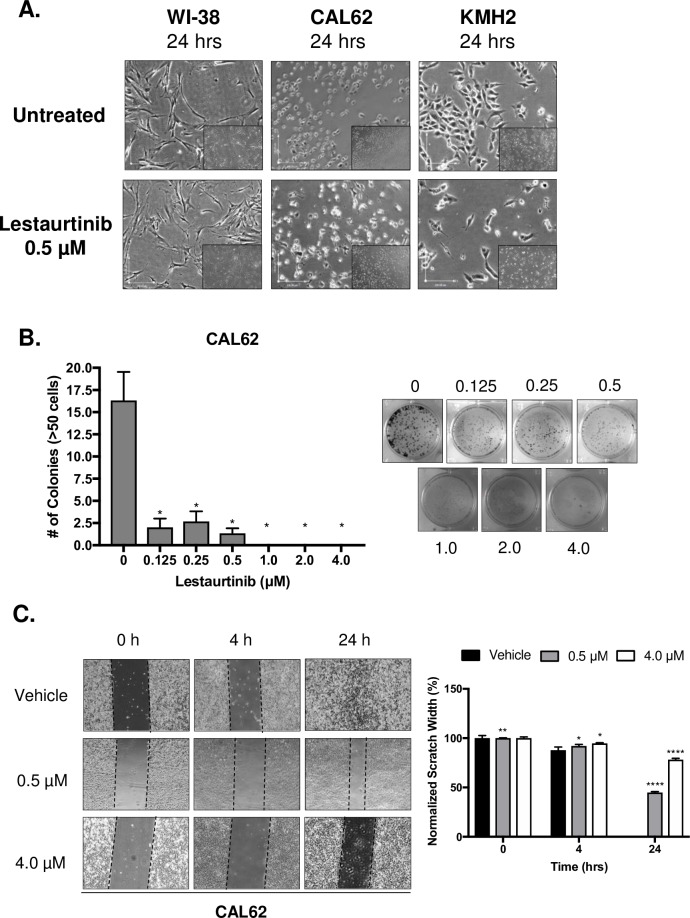
Fig 2. Treatment of ATC cell lines with Lestaurtinib resulted in an antiproliferative effect, in vitro.
(A) Phase contrast images of WI-38, CAL62 and KMH2 cell lines with and without Lestaurtinib treatment (0.5 μM; 4X magnification). (B) Clonogenic assay of CAL62 both untreated and treated with increasing concentrations of Lestaurtinib (0.125–4 μM) and stained with 0.5% crystal violet. Colony formation was quantified by counting colonies from 3 representative fields, with colonies defined as being ≥50 cells. (C) Scratch-wound assay of CAL62 cells treated with 0.5 and 4.0 μM Lestaurtinib at 0, 4 and 24 h. Ten measurements were taken across the width of the scratch and averaged (3 replicates per cell line) per time point. * represents p < 0.05, ** represents p < 0.01, *** represents p < 0.001, **** represents p < 0.0001, ns = not significant, Student’s unpaired, two-tail t-test.

