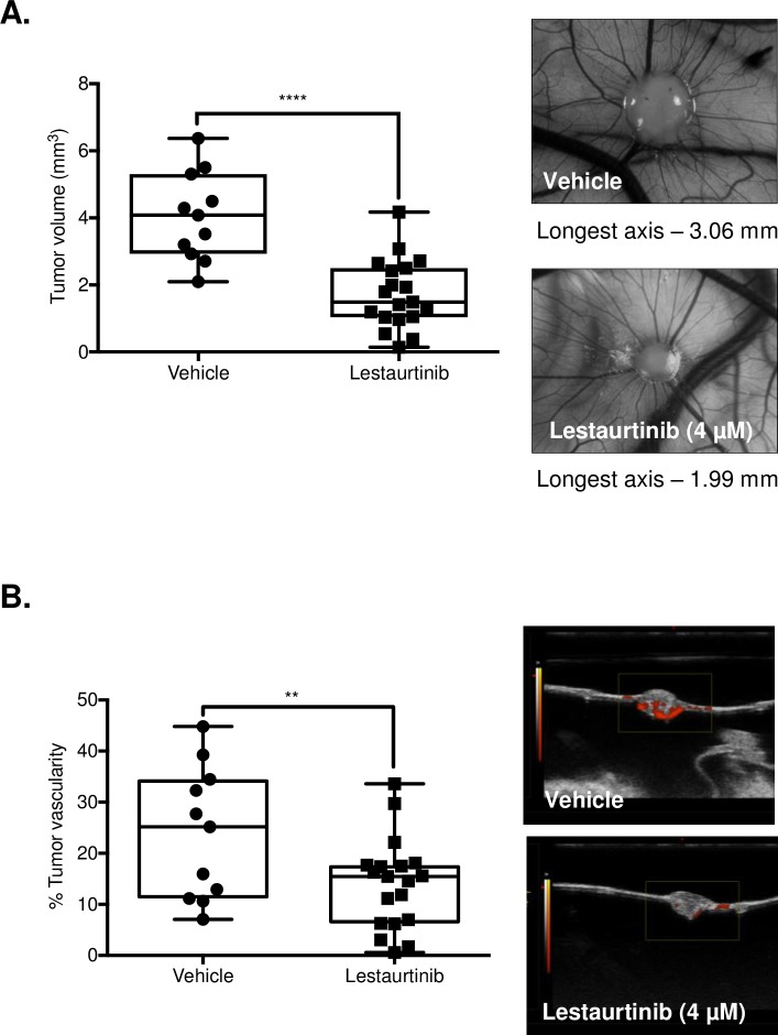
Fig 5. Lestaurtinib exhibits antiproliferative and antiangiogenic activity in the in vivo chick CAM membrane model.
(A) A total of 1 x 10⁶ cells per embryo were administered over top (onplant) of the chick embryo membrane, 9 days post-fertilization. Two days post-onplant, embryos were either treated with the vehicle alone or Lestaurtinib (4 μM), once daily for 5 days and sacrificed. Mean endpoint tumor volume was measured using ultrasound imaging in the vehicle-treated (n = 11) and Lestaurtinib-treated (n = 19) embryos. (B) Power Doppler ultrasound imaging measured the intergroup difference in fractional vascular volume at endpoint to generate mean percent vascularity between groups. * represents p < 0.05, ** represents p < 0.01, *** represents p < 0.001, **** represents p < 0.0001, ns = not significant, Student’s unpaired, two-tail t-test.

