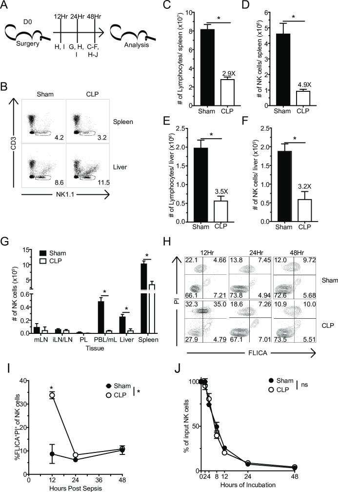
Fig 1. Apoptosis contributes to sepsis-induced systemic loss of NK-cells.
(A) Experimental Design. Sham or CLP mice were sacrificed 12, 24, or 48 hrs after surgery, and the number of NK-cells in the indicated tissues evaluated. (B) Representative flow plots of NK-cell gating. The total number of lymphocytes or NK-cells in spleen (C,D) or liver (E,F) 48 hrs after Sham or CLP surgery. (G) The number of NK-cells in mesenteric (mLN) and inguinal lymph nodes (iLN), peritoneal lavage (PL), blood (PBL/mL), liver, and spleen 24 hrs after sepsis-induction. (H) Representative flow plots of FLICA and PI staining of NK-cells. (I) Frequency of apoptotic (FLICA+PI+) NK-cells in the spleen at 12, 24, and 48 hrs after sepsis induction. (J) NK-cells obtained from Sham and CLP host 48 hrs post-surgery were placed in in vitro culture and the percent of surviving NK-cells was determined at indicated times. Data are representative from 3 independent experiments with 3–5 mice per group. Numbers above bars show fold change between groups. * p<0.05. Error bars represent the standard error of the mean.

