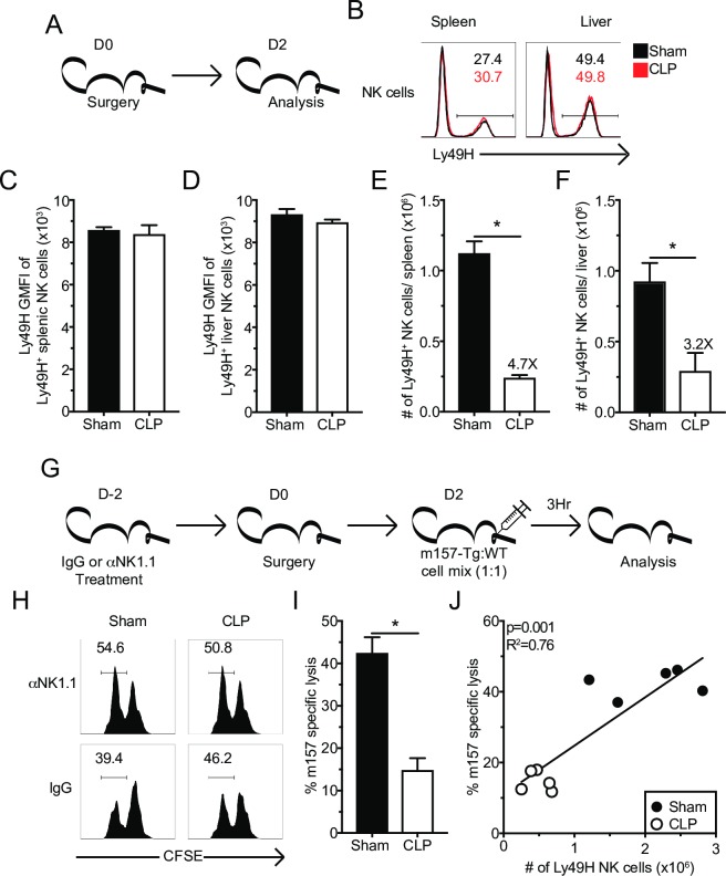
Fig 5. Sepsis results in numerical loss of Ly49H+ NK-cells and impaired in vivo killing.
(A) Experimental Design. Mice were sacrificed 2 days after sham or CLP surgery, and the number and Ly49H expression of Ly49H+ NK-cells in the spleen and liver determined. (B) Representative flow plots. Numbers indicate the frequency of Ly49+ NK-cells. The total number or GMFI of Ly49H by Ly49H+ NK-cells in spleen (C,E) or liver (D,F). (G) Experimental Design. Mice were treated with control IgG or α-NK1.1 depleting antibody prior to sepsis induction. Two days post-surgery all groups of mice received a 1:1 mixture of CFSE-labeled m157 expressing (m157-Tg) target (CFSElo) and m157-deficient littermate (WT) control (CFSEhi) cells. 3 hrs after injection mice were sacrificed and the ratio of m157-Tg to WT cells was determined. NK-depleted mice served as controls. (H) Representative flow plots. (I) m157 specific lysis in the spleen after Sham or CLP. (J) Correlation of m157 specific lysis with the number of Ly49H+ NK-cells in the spleen. Data are representative from 3 independent experiments with 3–5 mice per group. Numbers above bars show fold change between groups. * p<0.05. Error bars represent the standard error of the mean.

