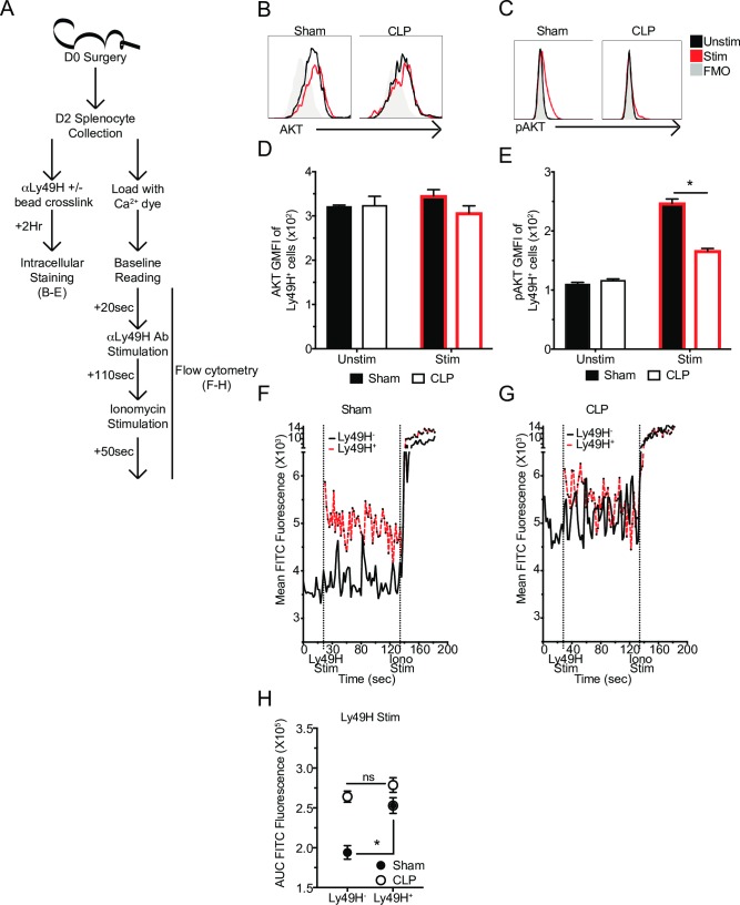
Fig 8. Reduced DAP12 expression is associated with defects in receptor signaling events.
(A) Experimental Design. Splenocytes were obtained 2 days post-surgery and were either labeled with α-Ly49H mAb and crosslinked with beads for 2 hrs and stained with AKT and pAKT antibodies or labeled with a calcium dye before being analyzed for calcium flux (baseline– 20 sec., Ly49H stim– 110 sec., Ionomycin– 50 sec.). Representative profiles or GMFI of AKT (B,D) or pAKT (C,E) in unstimulated and stimulated Ly49H+ NK-cells. Mean calcium dye fluorescence of Sham (F) or CLP (G) Ly49H+ or Ly49H- NK-cells during stimulation time course. (H) Area under the curve (AUC) for Ly49H- and Ly49H+ NK-cells during stimulation with α-Ly49H mAb. Data are representative from 2 independent experiments with 3 mice per group. * p<0.05. Error bars represent the standard error of the mean.

