
FIGURE 1.
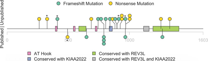
Schematic representation of mutations identified in 25 XGS patients that have participated in the XGS Registry. Mutations in five individuals who are not clinically assessed in detail, in this study, are indicated by an asterisk. Previously reported variants are indicated below the line, newly reported variants above. The x-axis shows amino acid positions in the encoded protein, pink rectangles indicate ATHook domains, green rectangles indicate REV3L homology domains, purple rectangle indicates KIAA2022 homology domains and gray rectangle indicates REV3L and KIAA2022 homology domains. Green circles indicate frameshift variants, yellow indicates nonsense variants (see Supplementary Material for further description of homologies and conservation)